Comparison of manual and automated fiber quantification tractography in patients with temporal lobe epilepsy

Abstract
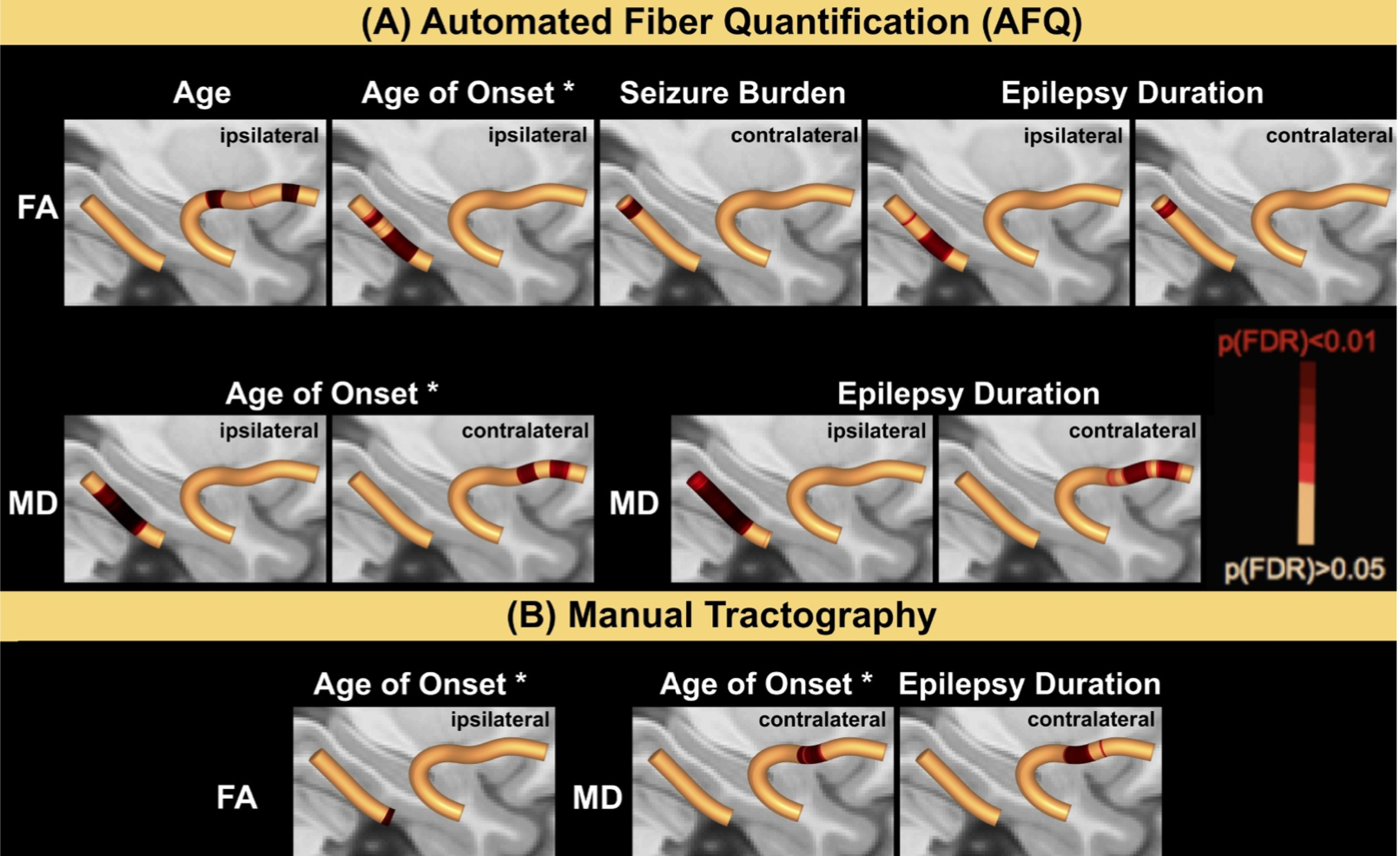
Objective: To investigate the agreement between manually and automatically generated tracts from diffusion tensor imaging (DTI) in patients with temporal lobe epilepsy (TLE). Whole and along-the-tract diffusivity metrics and cor- relations with patient clinical characteristics were analyzed with respect to tractography approach. Methods: We recruited 40 healthy controls and 24 patients with TLE who underwent conventional T1-weighted imaging and 60-direction DTI. An automated (Automated Fiber Quantification, AFQ) and manual (TrackVis) de- terministic tractography approach was used to identify the uncinate fasciculus (UF) and parahippocampal white matter bundle (PHWM). Tract diffusion scalar metrics were analyzed with respect to agreement across automated and manual approaches (Dice Coefficient and Spearman correlations), to side of onset of epilepsy and patient clinical characteristics, including duration of epilepsy, age of onset and presence of hippocampal sclerosis. Results: Across approaches the analysis of tract morphology similarity revealed Dice coefficients at moderate to good agreement (0.54 - 0.6) and significant correlations between diffusion values (Spearman's Rho=0.4–0.9). However, within bilateral PHWM, AFQ yielded significantly lower FA (left: Z = 4.4, p<0.001; right: Z = 5.1, p<0.001) and higher MD values (left: Z=-4.7, p<0.001; right: Z=-3.7, p<0.001) compared to the manual approach. Whole tract DTI metrics determined using AFQ were significantly correlated with patient char- acteristics, including age of epilepsy onset in FA (R = 0.6, p = 0.02) and MD of the ipsilateral PHWM (R=-0.6, p=0.02), while duration of epilepsy corrected for age correlated with MD in ipsilateral PHWM (R=0.7, p<0.01). Correlations between clinical metrics and diffusion values extracted using the manual whole tract technique did not survive correction for multiple comparisons. Both manual and automated along-the-tract analyses demonstrated significant correlations with patient clinical characteristics such as age of onset and epilepsy duration. The strongest and most widespread localized ipsi- and contralateral diffusivity alterations were observed in patients with left TLE and patients with HS compared to controls, while patients with right TLE and patients without HS did not show these strong effects. Conclusions: Manual and AFQ tractography approaches revealed significant correlations in the reconstruction of tract morphology and extracted whole and along-tract diffusivity values. However, as non-identical methods they differed in the respective yield of significant results across clinical correlations and group-wise statistics. Given the absence of excellent agreement between manual and AFQ techniques as demonstrated in the present study, caution should be considered when using AFQ particularly when used without reference to benchmark manual measures.